Sketch And Label Of A Cross Section Of A Long Bone : Bone Structure Anatomy And Physiology I
Sketch And Label Of A Cross Section Of A Long Bone : Bone Structure Anatomy And Physiology I. The outside of a bone is covered in a thin layer of dense irregular connective tissue called the periosteum. Terms in this set (3) epiphysis. A long bone is a bone that has greater length than width. Cartilaginous area at the ends of long bones where lengthwise growth takes place in the immature skeleton. This is an online quiz called long bone anatomy.
ads/bitcoin1.txt
What are the 2 kinds of bone marrow? The outside of a bone is covered in a thin layer of dense irregular connective tissue called the periosteum. Make a pencil sketch and use markers or colored pencils to add details. Draw and annotate the structure of a long bone 9 terms. 10% calcium carbonate (caco 3) 8.
Haversian Canal Wikipedia from upload.wikimedia.org The diaphysis is the tubular shaft that runs between the proximal and distal ends of the bone. The cut line is called a cutting plane, and can be done in several ways. The outside of a bone is covered in a thin layer of dense irregular connective tissue called the periosteum. 10% calcium carbonate (caco 3) 8. Cartilaginous area at the ends of long bones where lengthwise growth takes place in the immature skeleton. Create a drawing of the bone section in your laboratory journal and label the areas listed above. These are shown in localization. Please draw the distrabution of stresses for the cross section.
Sketch a cross section of an osteon and label its major parts.
ads/bitcoin2.txt
Sketch and label a cross section of a bone. A section view is a view used on a drawing to show an area or hidden part of an object by cutting away or removing some of that object. The clavicle is an extremely cutaneous bone. Calculate the stress at the tensile surface in terms of m if the inner radius r = 1.0 cm and the outer radius r = 1.5 cm. (do not copy and paste a picture from the text or internet.) The diaphysis is the tubular shaft that runs between the proximal and distal ends of the bone. Terms in this set (3) epiphysis. The diaphysis and the epiphysis. Please draw the distrabution of stresses for the cross section. Forms the larger rounded ends of long bones. What are the mineral crystals of bone called, and what are they made of? Cross section of long bone. Make a pencil sketch and use markers or colored pencils to add details.
A section view is a view used on a drawing to show an area or hidden part of an object by cutting away or removing some of that object. The diaphysis and the epiphysis. Looking at a bone in cross section, there are several distinct layered regions that make up a bone. It is the only long bone that is so superficial in the body and is very easily palpated. 4.1.1 label a diagram of a motor unit 6 terms.
Musculoskeletal System Anatomy And Functions Kenhub from thumbor.kenhub.com A long bone has a shaft and 2 ends. Cartilaginous area at the ends of long bones where lengthwise growth takes place in the immature skeleton. What is a section view ? The hollow region in the diaphysis is called the medullary cavity, which is filled. Make a pencil sketch and use markers or colored pencils to add details. In the space provided draw a longitudinal section of a long bone and label the following parte proximal epiphysis, distal epiphysis, diaphysis, metaphysis, medullary cavity, epiphyseal line 2. Consider a long bone under bending and assume that the bone can be modeled as a hollow cylindrical tube. Draw and label the following structures as they appear using the 10x objective o bone marrow o bony trabeculae 27.
Plates of cartilage, also known as growth plates which allow the long bones to grow during childhood.
ads/bitcoin2.txt
Continue to label this drawing as you explore the inside of the bone. Looking at a bone in cross section, there are several distinct layered regions that make up a bone. Create a drawing of the bone section in your laboratory journal and label the areas listed above. A long bone is a bone that has greater length than width. 3d diagram of long bone 12 photos of the 3d diagram of long bone , bone. What is a section view ? Forms the larger rounded ends of long bones. These are shown in localization. The outside of a bone is covered in a thin layer of dense irregular connective tissue called the periosteum. A section view is a view used on a drawing to show an area or hidden part of an object by cutting away or removing some of that object. The original can be viewed here: Growth in length of a bone occurs at the 4. A long bone has a shaft and 2 ends.
Draw and annotate the structure of a long bone 9 terms. The ends of a long bone contain spongy bone and an epiphyseal line. There is a printable worksheet available for download here so you can take the quiz with pen and paper. 4.1.1 label a diagram of a motor unit 6 terms. A cross section of a human long bone.
The Structure Of A Long Bone Humerus Ppt Video Online Download from slideplayer.com Continue to label this drawing as you explore the inside of the bone. Looking at a bone in cross section, there are several distinct layered regions that make up a bone. 4.1.1 label a diagram of a motor unit 6 terms. Calculate the stress at the tensile surface in terms of m if the inner radius r = 1.0 cm and the outer radius r = 1.5 cm. The periosteum contains many strong collagen fibers that are used to firmly anchor tendons and muscles to the bone for movement. Bone matrix and cells bone matrix osseous tissue is a connective tissue and like all connective tissues contains relatively few cells and large amounts of extracellular matrix. Bone anatomy of the neck (do not copy and paste a picture from the text or internet.)
Growth in length of a bone occurs at the 4.
ads/bitcoin2.txt
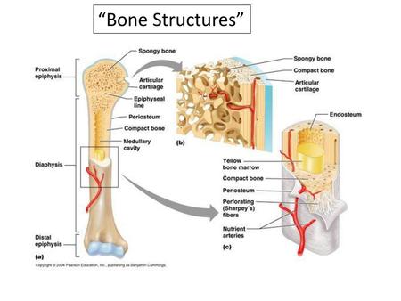
Continue to label this drawing as you explore the inside of the bone. The hollow region in the diaphysis is called the medullary cavity, which is filled with yellow marrow. Please draw the distrabution of stresses for the cross section. Sketch and label a cross section of a bone. The cut line is called a cutting plane, and can be done in several ways. Draw and label the following structures as they appear using the 10x objective o bone marrow o bony trabeculae 27. The ends of a long bone contain spongy bone and an epiphyseal line. A = epiphysis b = diaphysis c = articular cartilage d = periosteum f = compact bone g = medullary cavity (yellow marrow) h = endosteum j = epiphyseal line (growth plate) coloring worksheet for this image. Sketch a cross section of an osteon and label its major parts. Label the parts of a long bone. A long bone is a bone that has greater length than width. It is the first bone to ossify in the fetus from membrane, and it does so from 2 centres, which ossify at the 5th week and rapidly fuse in the midline. Click on the tags below to find other quizzes on the same subject.
ads/bitcoin3.txt
ads/bitcoin4.txt
ads/bitcoin5.txt
0 Response to "Sketch And Label Of A Cross Section Of A Long Bone : Bone Structure Anatomy And Physiology I"


0 Response to "Sketch And Label Of A Cross Section Of A Long Bone : Bone Structure Anatomy And Physiology I"
Post a Comment